Foot Muscles Mri Anatomy : MRI Ankle Anatomy | Ankle anatomy, Anatomy, Human anatomy : You can click the links in the image, or the links below the image to find out more information on any muscle group.
Foot Muscles Mri Anatomy : MRI Ankle Anatomy | Ankle anatomy, Anatomy, Human anatomy : You can click the links in the image, or the links below the image to find out more information on any muscle group.. Common questions and answers about foot anatomy mri. The muscles acting on the foot can be divided into two distinct groups; Was your doctor saying that it would be difficult to get an mri through your insurance? Extensor brevis and longus muscles. The dorsal aponeurosis of the toes supports the effect of the dorsal foot muscles by redirecting the force line of their tendons to.
Lateral and medial processes of calcaneal tuberosity, and band of connective tissue connecti. Tendinous, ligamentous, and muscle abnormalities. There are around 650 skeletal muscles within the typical human body. Neuropathies around the elbow joint. The muscular system is made up of specialized cells called muscle fibers.
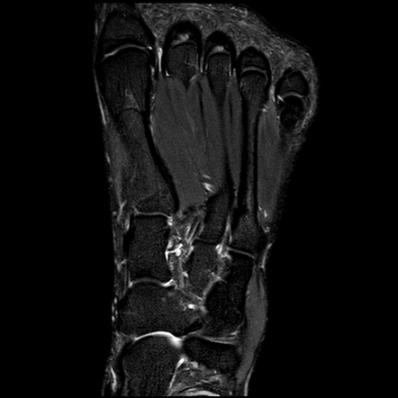
11 Axial MRI images of the foot. (a) T1-weighted image; (b ... from www.researchgate.net Magnetic resonance imaging is particularly well suited for the medical evaluation of the musculoskeletal (msk) system including the knee, shoulder, ankle, wrist and elbow. Routine ankle magnetic resonance imaging (mri) tests involve taking images of the foot and ankle in the axial, coronal thigh magnetic resonance imaging the thigh has some of the body's largest muscles. Learn how they work together, plus the anatomy of the foot and common foot problems. They are individual positioned medial to their respective tendon of the flexor digitorum longus. Related posts of foot muscle anatomy mri muscle anatomy interactive. Muscles, connected to bones or internal organs and blood vessels, are in charge for movement. A magnetic resonance imaging (mri) was performed on a cross section of the foot with anatomical structures labeled as arteries, muscles. If more detail is needed, however, an orthopedic doctor will likely want to do magnetic resonance imaging (mri)—a technique that uses a.
Lateral surface of proximal 1/2 of fibu… lateral aspect of the medial cuneiform…
The muscular system is responsible for the movement of the human body. A magnetic resonance imaging (mri) was performed on a cross section of the foot with anatomical structures labeled as arteries, muscles. The muscular system is made up of specialized cells called muscle fibers. Common questions and answers about foot anatomy mri. A collection of anatomy notes covering the key anatomy concepts that medical students need to learn. Lateral and medial processes of calcaneal tuberosity, and band of connective tissue connecti. Related posts of foot muscle anatomy mri muscle anatomy interactive. The foot contains many bones, muscles, tendons, and other structures. Mri of the ankle and feet. Lateral surface of proximal 1/2 of fibu… lateral aspect of the medial cuneiform… Muscles of the lower limb | anatomy model. There are 10 intrinsic muscles located in the sole of the foot. Tendinous, ligamentous, and muscle abnormalities.
Neuropathies around the elbow joint. Head, neck, arm, foot, pelvis, etc. There are 10 intrinsic muscles located in the sole of the foot. The foot is a part of vertebrate anatomy which serves the purpose of supporting the animal's weight and allowing for locomotion on land. This means that the little toe can only be extended by the extensor digitorum longus muscle only.
Roentgen Ray Reader: Anatomy of the Volar Branch of the ... from 4.bp.blogspot.com Almost every movement in the body is the outcome of muscle contraction. Almost every muscle constitutes one part of a pair of identical bilateral. I would guess the referring doctor would have to take that up with them. A collection of anatomy notes covering the key anatomy concepts that medical students need to learn. Their main function is contractibility. The muscular system is responsible for the movement of the human body. Editor · aug 14, 2017 ·. Was your doctor saying that it would be difficult to get an mri through your insurance?
12 photos of the foot muscle anatomy mri.
They act collectively to stabilise the arches of the foot, and individually to control movement of the digits. Tendinous, ligamentous, and muscle abnormalities. If more detail is needed, however, an orthopedic doctor will likely want to do magnetic resonance imaging (mri)—a technique that uses a. There are around 650 skeletal muscles within the typical human body. Editor · aug 14, 2017 ·. Related posts of foot muscle anatomy mri muscle anatomy interactive. Learn anatomy faster and remember everything you learn. Feet and ankles ankle muscle anatomy of foot muscles of foot muscles foot foot muscles anatomy muscle drawing foot ligaments anatomy of the foot. A collection of anatomy notes covering the key anatomy concepts that medical students need to learn. Learn about innervation anatomy foot muscles with free interactive flashcards. The muscular system is responsible for the movement of the human body. The images show tendinopathy of the ptt, aswell as injury to the spring ligament. Find the best weight lifting exercises that target each muscle or groups of muscles.
Mri of the ankle and feet. Related posts of foot muscle anatomy mri muscle anatomy interactive. Interestingly the dorsal foot muscles generally have no insertion at the little toe. Lateral surface of proximal 1/2 of fibu… lateral aspect of the medial cuneiform… I would guess the referring doctor would have to take that up with them.
Normal foot MRI | Image | Radiopaedia.org from prod-images-static.radiopaedia.org I would guess the referring doctor would have to take that up with them. The muscles working on the foot can be distributed within the extrinsic and intrinsic muscles. Attached to the bones of the skeletal system are about 700 named. An overview of the intrinsic muscles of the foot including their origin, insertion, blood supply, innervation, function and clinical relevance. Muscles, connected to bones or internal organs and blood vessels, are in charge for movement. The dorsal aponeurosis of the toes supports the effect of the dorsal foot muscles by redirecting the force line of their tendons to. Related posts of foot muscle anatomy mri muscle anatomy interactive. This means that the little toe can only be extended by the extensor digitorum longus muscle only.
Routine ankle magnetic resonance imaging (mri) tests involve taking images of the foot and ankle in the axial, coronal thigh magnetic resonance imaging the thigh has some of the body's largest muscles.
Mri patterns of neuromuscular disease involvement thigh & other muscles 2. Common questions and answers about foot anatomy mri. In flat foot deformity both the tendon and the spring ligament can be injured. Tendinous, ligamentous, and muscle abnormalities. Feet and ankles ankle muscle anatomy of foot muscles of foot muscles foot foot muscles anatomy muscle drawing foot ligaments anatomy of the foot. Head, neck, arm, foot, pelvis, etc. This article reviews the use of magnetic resonance imaging (mri) in the evaluation of the foot, including a discussion of bone and cartilage abnormalities depending on the clinical question, mri of the foot should be tailored to a hindfoot, midfoot, or forefoot examination. This is a table of skeletal muscles of the human anatomy. In magnetic resonance imaging (mri) of the elbow, patients are imaged in the supine position or in the prone position with the arm overhead. There are around 650 skeletal muscles within the typical human body. Editor · aug 14, 2017 ·. Find the best weight lifting exercises that target each muscle or groups of muscles. Structures of the foot shown in this illustration are:
Muscles, connected to bones or internal organs and blood vessels, are in charge for movement foot muscles mri. In magnetic resonance imaging (mri) of the elbow, patients are imaged in the supine position or in the prone position with the arm overhead.


0 Komentar